Rib Cage Anatomy / Rib Cage Wikipedia
The ribs are a veritable collection of bone, muscle, and organs, most of which are fairly important for living and other useful functions. The thoracic cage consists of the 12 thoracic vertebrae, the associated intervertebral discs, 12 pairs of ribs with their costal cartilages, and the sternum. The thoracic cage takes the form of a domed bird cage with the horizontal bars formed by ribs and costal cartilages. Together with the sternum, thoracic vertebrae, and costal cartilages, the ribs form the thoracic cage, also called the bony thorax. Learn more about the skeletal system with quizzes and labelling exercises. Each pair is numbered based on their attachment to the sternum, a bony process at the front of the rib cage which serves as an anchor point. It may occur after an obvious injury or without explanation. Muscle anatomy study guide 12 photos of the muscle anatomy study guide anatomy and physiology muscle study guide, anatomy physiology muscle study guide, cat muscle anatomy study guide, muscle anatomy study guide, muscle study guide for anatomy, human muscles, anatomy and physiology muscle study guide, anatomy physiology. Rib cage anatomy and its implications in back pain 'it is important to understand rib cage anatomy if we want to treat upper back pain' explains sarah key. The top edge of the manubrium has a depression called the suprasternal or jugular notch. It is formed by the 12 thoracic vertebrae, 12 pairs of ribs and associated costal cartilages and the sternum.
There are twelve (12) pairs of ribs and all articulate posteriorly with the thoracic vertebrae. It is supported by the vertical sternum or breastbone (anteriorly) and the 12 thoracic vertebrae. The first rib is right up in the angle of the neck and shoulder. Each pair is numbered based on their attachment to the sternum, a bony process at the front of the rib cage which serves as an anchor point. As part of the bony thorax, the ribs protect the internal thoracic organs. Introduction to the structure of the ribcage and ribs: On the interior wall of the rib body is a channel, sulcus costae, with blood vessels and nerves. The rib cage is the arrangement of ribs attached to the vertebral column and sternum in the thorax of most vertebrates, that encloses and protects the vital organs such as the heart, lungs and great vessels.
It is made up of 12 pairs of ribs.
The ribs are attached to the breastbone, which is the. Check out our anatomy rib cage art selection for the very best in unique or custom, handmade pieces from our shops. Ninja nerds!join us in this video where we show the sternum and rib articulation anatomy through the use of a model. Elevates the ribs, increasing the thoracic volume. Anatomy the rib cage is a bony structure found in the chest (thoracic cavity). The first rib is right up in the angle of the neck and shoulder. There are 11 pairs of external intercostal muscles. However, pain can mean different things depending on location and other associated symptoms. There are twelve (12) pairs of ribs and all articulate posteriorly with the thoracic vertebrae. There are twelve pairs of ribs, all of which articulate with the vertebral column. They articulate with the vertebral column posteriorly, and terminate anteriorly as cartilage (known as costal cartilage). Although that is one key function, the ribcage does so much more. Rib cage anatomy labeled vector illustration diagram medical human chest skeletal bone structure model numbered ribs sternum cartilage parts and clavicular articulation health care education premium vector in adobe illustrator. Lessons on the bone markings of the ribs and sternum. It may occur after an obvious injury or without explanation.
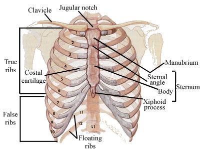
Rib cage pain may be sharp, dull, or achy and felt at or below the chest or above the navel on either side. Anatomy the rib cage has 12 sets of ribs. The ribs protect vital organs within the thoracic cage, and they also assist. There are twelve pairs of ribs, all of which articulate with the vertebral column. The ribs are a veritable collection of bone, muscle, and organs, most of which are fairly important for living and other useful functions. Originate at the lower border of the rib, inserting into the superior border of the rib below. Rib cage anatomy the rib cage, shaped in a mild cone shape and more flexible than most bone sets, is made up of varying elements such as the thoracic vertebra, 12 equally paired ribs, costal cartilage, and held together anteriorly by the sternum. The sternum is a flat bone that is made up of three parts, the (1) manubrium, (2) body, and the (3) xiphoid process.

However, pain can mean different things depending on location and other associated symptoms.
There are 11 pairs of external intercostal muscles. It may occur after an obvious injury or without explanation. Introduction to the structure of the ribcage and ribs: Although that is one key function, the ribcage does so much more. Anatomy the rib cage is a bony structure found in the chest (thoracic cavity). It is made up of 12 pairs of ribs. It is formed by the 12 thoracic vertebrae, 12 pairs of ribs and associated costal cartilages and the sternum. The sternum is a flat bone that is made up of three parts, the (1) manubrium, (2) body, and the (3) xiphoid process. The bones of the rib cage are the sternum, the 12 thoracic vertebrae and the 12 pairs of ribs. The thoracic cage consists of the 12 thoracic vertebrae, the associated intervertebral discs, 12 pairs of ribs with their costal cartilages, and the sternum. Check out our anatomy rib cage art selection for the very best in unique or custom, handmade pieces from our shops. Ninja nerds!join us in this video where we show the sternum and rib articulation anatomy through the use of a model. The ribs protect vital organs within the thoracic cage, and they also assist.
The ribs are a veritable collection of bone, muscle, and organs, most of which are fairly important for living and other useful functions. Rib cage pain can be caused. Embroidery human rib cage with red roses. Anatomy the rib cage has 12 sets of ribs. Contributing to their role in protecting the internal thoracic organs.

The cervicothoracic junction is where the neck (cervical spine) connects with the upper back (thoracic spine).
The ribs protect vital organs within the thoracic cage, and they also assist. The ribs are curved, flat bones which form the majority of the thoracic cage. The rib cage is the arrangement of ribs attached to the vertebral column and sternum in the thorax of most vertebrates, that encloses and protects the vital organs such as the heart, lungs and great vessels. The first rib is right up in the angle of the neck and shoulder. Related posts of muscle anatomy rib cage muscle anatomy study guide. The ribs are a set of twelve paired bones which form the protective 'cage' of the thorax. Rib cage pain can be caused. It is made up of 12 pairs of ribs. Consequently, pain from the rib cage tends to cause alarm, especially if it comes on suddenly. As viewed from the side, the thoracic spine's vertebrae form a kyphotic curve that runs from t1 to t12, in which the spine curves outward towards the back of the body to allow more room for the internal organs such. They run inferoanteriorly from the rib above to the rib below, and are continuous with the external oblique of the abdomen.

Originate at the lower border of the rib, inserting into the superior border of the rib below.

The ribs are curved, flat bones which form the majority of the thoracic cage.

They run inferoanteriorly from the rib above to the rib below, and are continuous with the external oblique of the abdomen.

Consequently, pain from the rib cage tends to cause alarm, especially if it comes on suddenly.

A rib has a flat body, as you can see from the picture of the anatomy of the human rib cage.

The ribs are curved, flat bones which form the majority of the thoracic cage.

It is supported by the vertical sternum or breastbone (anteriorly) and the 12 thoracic vertebrae.
Ninja nerds!join us in this video where we show the sternum and rib articulation anatomy through the use of a model.

The thoracic cage (rib cage) is the skeletal framework of the thoracic wall, which encloses the thoracic cavity.
 manubrium, (2) body, and the (3) xiphoid process.")
It is supported by the vertical sternum or breastbone (anteriorly) and the 12 thoracic vertebrae.

Rib cage anatomy labeled vector illustration diagram medical human chest skeletal bone structure model numbered ribs sternum cartilage parts and clavicular articulation health care education premium vector in adobe illustrator.
They run inferoanteriorly from the rib above to the rib below, and are continuous with the external oblique of the abdomen.

There are 11 pairs of external intercostal muscles.
 pairs of ribs and all articulate posteriorly with the thoracic vertebrae.")
The thoracic cage (rib cage) is the skeletal framework of the thoracic wall, which encloses the thoracic cavity.

Lateral view of a pair of ribs articulating with the thoracic vertebrae.

Originate at the lower border of the rib, inserting into the superior border of the rib below.

Check out our anatomy rib cage art selection for the very best in unique or custom, handmade pieces from our shops.

The cervicothoracic junction is where the neck (cervical spine) connects with the upper back (thoracic spine).

As part of the bony thorax, the ribs protect the internal thoracic organs.

Instead, anatomists classify the ribs as flat bones, and they are located within the axial skeleton.

Rib cage anatomy and its implications in back pain 'it is important to understand rib cage anatomy if we want to treat upper back pain' explains sarah key.
 is the skeletal framework of the thoracic wall, which encloses the thoracic cavity.")
Rib cage anatomy labeled vector illustration diagram medical human chest skeletal bone structure model numbered ribs sternum cartilage parts and clavicular articulation health care education premium vector in adobe illustrator.

The ribs are a veritable collection of bone, muscle, and organs, most of which are fairly important for living and other useful functions.

The ribs are a set of twelve paired bones which form the protective 'cage' of the thorax.

A cervical rib forms from the overdevelopment of the transverse process of a cervical vertebra, typically from the seventh cervical vertebra in the neck known as c7.

They are extremely light, but highly resilient;

However, only seven have a direct articulation with the sternum.

Rib cage anatomy and its implications in back pain 'it is important to understand rib cage anatomy if we want to treat upper back pain' explains sarah key.

The thoracic cage takes the form of a domed bird cage with the horizontal bars formed by ribs and costal cartilages.

Elevates the ribs, increasing the thoracic volume.

There are twelve pairs of ribs, all of which articulate with the vertebral column.

Lateral view of a pair of ribs articulating with the thoracic vertebrae.

Rib cage anatomy the rib cage, shaped in a mild cone shape and more flexible than most bone sets, is made up of varying elements such as the thoracic vertebra, 12 equally paired ribs, costal cartilage, and held together anteriorly by the sternum.

Rib cage anatomy and its implications in back pain 'it is important to understand rib cage anatomy if we want to treat upper back pain' explains sarah key.

Posting Komentar untuk "Rib Cage Anatomy / Rib Cage Wikipedia"